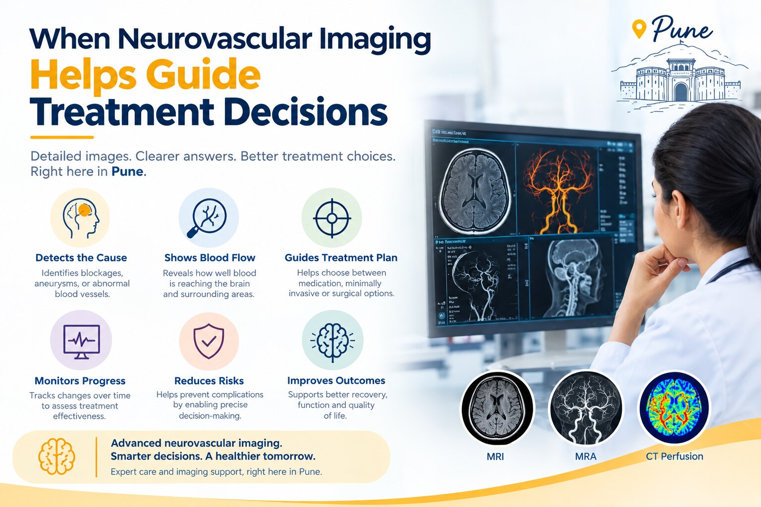
A sudden neurological symptom can require imaging within an emergency treatment window, while other vascular findings need carefully sequenced scans before anyone recommends an intervention. By the end, you will know which scan answers which question, how findings affect treatment choices, and what to ask about interpretation, urgency and next steps in Pune.
Key takeaways
- Treat sudden neurological symptoms as an emergency, not an outpatient imaging request.
- CT and MRI show different brain injuries; angiography maps vessels for treatment planning.
- Imaging helps specialists choose between medicines, thrombectomy, coiling, clipping and venous procedures.
- Ask whether treatment-planning images show the lesion, surrounding anatomy and blood-flow pattern clearly.
When a neurological symptom calls for immediate imaging
Sudden weakness or numbness on one side, trouble speaking, new vision loss, a thunderclap headache, seizure, collapse, or reduced alertness requires emergency assessment and neurovascular imaging rather than an outpatient appointment.
The first decision is whether the symptom signals intracranial bleeding, an arterial blockage, or another time-critical process, because each pathway demands different imaging and treatment.
| Symptom pattern | Why it cannot wait | Initial imaging question |
|---|---|---|
| Sudden weakness, facial droop, or speech trouble | Possible acute ischaemic stroke or haemorrhage | Does non-contrast CT show bleeding, and does CTA show a large-vessel blockage? |
| Sudden severe headache, vomiting, neck stiffness, or collapse | Possible subarachnoid haemorrhage or aneurysm rupture | Is there blood on CT, and is an aneurysm visible on CTA? |
| New seizure, confusion, or loss of consciousness | Possible bleeding, venous thrombosis, tumour, or stroke | Is urgent CT enough, or are venous imaging or MRI needed? |
| Sudden vision loss or double vision | Possible retinal, carotid, posterior-circulation, or brain ischaemia | Is an arterial blockage or dissection present? |
For suspected acute stroke, non-contrast CT is usually the first decisive scan; a normal early result does not exclude ischaemia. CTA follows when a treatable arterial occlusion, dissection, aneurysm, or carotid problem is suspected.
Understanding when neurovascular imaging is used in Pune also means balancing kidney function, prior iodinated-contrast reactions, pregnancy when relevant, and the ability to lie still against the danger of delaying vessel imaging. MRI is not automatically the safest first test during an unstable, hyperacute presentation.
What each scan can reveal about the brain and its blood vessels
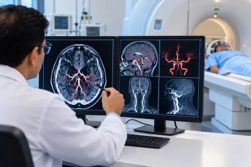
No single scan answers every neurovascular question: each shows different tissue, vessels or blood-flow behaviour.
| Scan | What it can reveal | When it helps |
|---|---|---|
| Non-contrast CT | Haemorrhage, mass effect and established infarction | First test for suspected acute stroke; a normal early result does not exclude ischaemia |
| CT angiography (CTA) | Arterial blockage, stenosis, aneurysm and some AVMs | Locates a large-vessel occlusion and supports thrombectomy or aneurysm planning |
| CT venography (CTV) | Contrast-filled cerebral veins and venous sinuses | Confirms cerebral venous sinus thrombosis and related venous infarction or haemorrhage |
| CT perfusion | Blood flow, blood volume and delayed transit; estimates core and salvageable tissue | Helps select patients for reperfusion treatment |
| MRI | Acute infarction, small haemorrhage, oedema and tissue detail | Clarifies lesions that CT misses and assesses AVM-related injury |
| MRA | Cerebral arteries without catheterisation | Screens or follows aneurysms and arterial narrowing |
| MR venography | Cerebral venous drainage | Detects venous sinus occlusion without ionising radiation |
| Doppler ultrasound | Blood-flow speed and direction in neck or selected brain vessels | Assesses carotid stenosis and monitors flow |
| Catheter cerebral angiography (DSA) | Detailed arteries, feeders, drainers, fistulas and flow | Maps lesions before embolisation, surgery or radiosurgery; it carries procedural risks |
| Spinal angiography | Arteries and veins supplying the spinal cord | Locates spinal AVMs or dural fistulas before treatment |
Contrast choice, kidney function and prior reactions affect planning. Neurovascular imaging applications in Pune are most valuable when findings change treatment, not when another scan merely repeats an answer.
How imaging findings change stroke, aneurysm and venous-treatment decisions
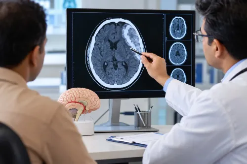
A negative non-contrast CT does not rule out an ischaemic stroke. If CT shows no haemorrhage, CT angiography can identify an accessible large-vessel blockage, while CT perfusion or MRI diffusion/perfusion can estimate irreversibly injured core versus salvageable tissue.
These findings determine whether urgent thrombolysis assessment or thrombectomy is appropriate; selected patients qualify for thrombectomy up to 6–24 hours from last known well, based on clinical findings, vessel location, infarct core and pre-stroke function.
- Stroke: No haemorrhage plus a treatable large-vessel occlusion supports thrombectomy assessment. A large established infarct, inaccessible vessel or absent salvageable tissue may shift the plan to medical management.
- Aneurysm: CTA, MRA or catheter angiography shows the aneurysm’s neck, size, branches and rupture pattern. Those details guide observation, coiling, flow diversion, surgery or another approach.
- Venous sinus thrombosis: CT venography or MR venography confirms the blocked sinus and shows venous infarction or haemorrhage. Anticoagulation remains central; deterioration despite treatment can prompt endovascular rescue review.
- AVM or fistula: Catheter angiography maps feeding arteries, drainage veins and high-risk features before embolisation, surgery or radiosurgery is considered.
Advanced brain vessel imaging in Pune is useful only when it reaches the decision-maker quickly. Teams also weigh kidney function, prior iodinated-contrast reactions, pregnancy when relevant, ability to lie still and access to intervention, anaesthesia and transfer. A scan software score never replaces the examination and specialist review.
What treatment planning images must show before an intervention
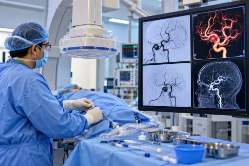
Before choosing an intervention, imaging must show the lesion’s exact location, size, shape, blood supply, drainage and relationship to normal brain and arteries. A neurovascular scan for treatment planning in Pune must also reveal whether the anatomy makes treatment safer than observation.
| Treatment | Images must establish | Why it matters |
|---|---|---|
| Coiling | Aneurysm dome, neck width, parent artery, branch vessels and perforators | A wide neck or an artery arising from the sac can require balloon or stent assistance, surgery or another approach. |
| Flow diversion | Parent-vessel diameter, landing zones, side branches, perforators and nearby aneurysms | The device must cover the neck without blocking essential branches or leaving inadequate vessel contact. |
| AVM embolisation | Every feeding artery, nidus compartment, fistulous point, draining vein, flow pattern and associated aneurysm | An unrecognised feeder or premature venous occlusion can cause infarction or haemorrhage. |
| Surgery | Lesion depth, access corridor, relation to eloquent cortex, cranial nerves and major vessels | Surgeons need a safe route and must know what normal tissue lies between the opening and target. |
| Radiosurgery | Nidus volume, margins, location and distance from optic pathways, brainstem and other critical structures | Dose and targeting depend on precise three-dimensional boundaries. |
MRI or CT must also show haemorrhage, oedema, infarction and mass effect. Catheter angiography supplies the dynamic detail when non-invasive scans cannot map small feeders, flow or venous drainage; if those details remain uncertain, choosing treatment is premature.
From emergency scan to specialist decision in Pune
After the first scan, an emergency radiologist reviews the images with the treating stroke physician. For suspected large-vessel stroke, non-contrast CT looks for haemorrhage and established infarction; CT angiography then checks for an arterial blockage. A normal early CT does not rule out ischaemic stroke.
The next decision depends on what remains uncertain and what treatment is available:
- CT perfusion or MRI diffusion/perfusion imaging can estimate salvageable tissue when onset is unknown or presentation is late. The software result is checked against the examination, vessel scan, infarct core and pre-stroke disability.
- MRI is useful for small infarcts, posterior-fossa stroke and stroke mimics, but it should not delay reperfusion treatment in an unstable patient.
- Catheter angiography is chosen when detailed vessel mapping could change treatment, such as aneurysm occlusion, AVM embolisation or fistula repair.
- CT venography or MR venography is added when cerebral venous thrombosis remains a concern.
Contrast planning includes kidney function, previous iodinated-contrast reactions, pregnancy status when relevant, and the ability to lie still. In suspected large-vessel stroke, a modest creatinine abnormality must be weighed against treatment delay.
Advanced brain vessel imaging in Pune works best when a neuroradiologist, neurointerventionalist, neurologist, anaesthetist and surgeon can act on the result. Dr Santosh Patil-Endovascular Neuro Surgeon Pune can fit into this pathway when specialist interpretation, procedure planning or rapid transfer is needed beyond the first facility’s capability.
Related service
Neurovascular Imaging What is Neurovascular Imaging- Neurovascular imaging plays a crucial role in the diagnosis, characterization, and treatment planning of the... View service → |
Frequently asked questions
When is neurovascular imaging used in Pune?
It is used urgently for sudden weakness, speech trouble, vision loss, thunderclap headache, seizure, collapse or reduced alertness. Specialists also use it to investigate suspected aneurysms, stroke and venous disorders.
What can CT, MRI and angiography reveal about the brain and its blood vessels?
CT can rapidly show bleeding and major structural changes. MRI can reveal early or small areas of brain injury. CT angiography, MR angiography and catheter angiography map vessel narrowing, blockage, aneurysms and abnormal connections.
How do imaging findings change stroke, aneurysm and venous-treatment decisions?
Findings help determine whether stroke treatment involves medication or thrombectomy, whether an aneurysm needs observation or repair, and whether a venous disorder requires anticoagulation, endovascular treatment or another approach.
What must treatment-planning images show before an intervention?
They must identify the target lesion, its size and location, nearby branches, the affected vessel, blood-flow direction and access anatomy. The treating team also needs images clear enough to select the safest procedure.
Keywords