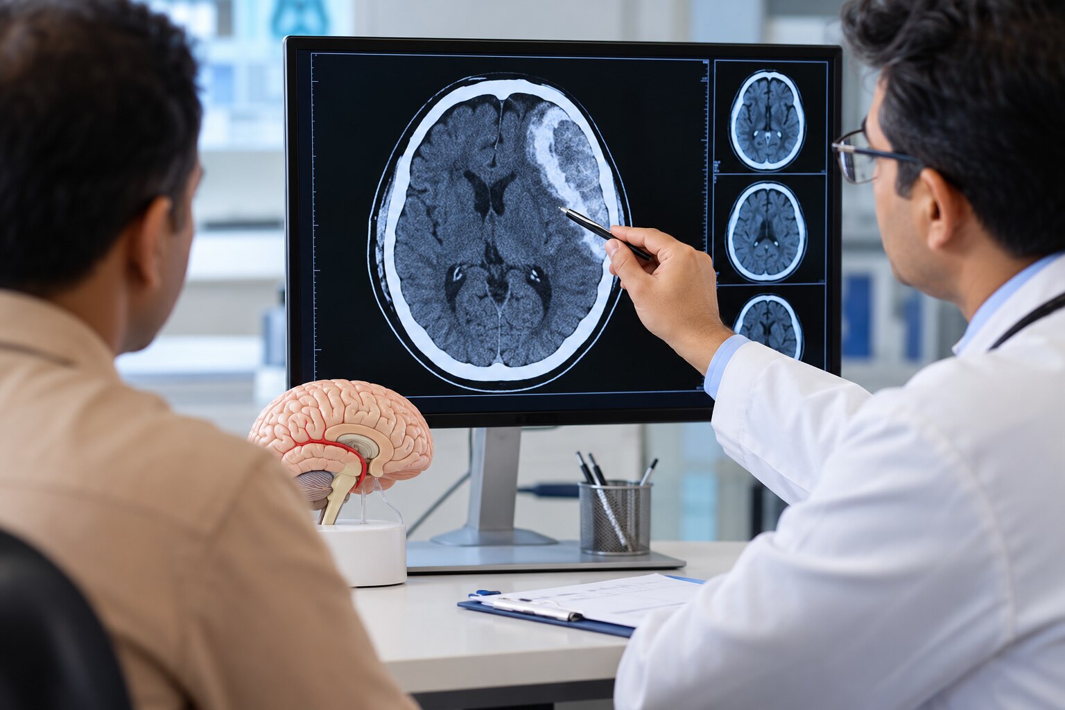
A subdural bleed needs urgent specialist assessment when symptoms worsen, the CT shows significant compression, or the patient has risk factors that make expansion more likely. By the end, you will know which warning signs require hospital care, how acute and chronic bleeds differ, and when observation, surgery, or endovascular treatment may be considered.
Key takeaways
- Seek emergency care for worsening headache, vomiting, drowsiness, weakness, seizures, or unequal pupils.
- CT findings such as brain compression and midline shift can make treatment urgent.
- Observation requires neurological checks, medication review, and repeat CT imaging.
- Burr-hole drainage, craniotomy, and embolization serve different clinical situations.
Which symptoms and CT findings make treatment urgent?
A worsening headache, repeated vomiting, increasing drowsiness, confusion, weakness on one side, slurred speech, a seizure, unequal pupils, or loss of consciousness requires emergency hospital assessment. Do not wait for a routine appointment: these signs can indicate brain compression or rising intracranial pressure after a fall, injury, or bleeding while taking anticoagulant or antiplatelet medicine.
For anyone asking “when does subdural hemorrhage need treatment in Pune,” the answer depends on both the neurological examination and the non-contrast CT head. In an acute traumatic subdural hematoma, commonly used surgical thresholds are clot thickness above 10 mm or midline shift above 5 mm, regardless of the Glasgow Coma Scale score.
A smaller collection is not automatically safe. Urgent neurosurgical treatment may still be needed when the patient’s Glasgow Coma Scale falls by at least 2 points, pupils become unequal or abnormal, neurological function worsens, or intracranial pressure exceeds 20 mm Hg.
| Finding | What it means | Action |
|---|---|---|
| Worsening symptoms | The brain may be under increasing pressure | Go to emergency care immediately |
| More than 10 mm clot thickness | Large acute traumatic hematoma | Urgent neurosurgical assessment |
| More than 5 mm midline shift | The brain has moved from its normal position | Urgent surgical consideration |
| Smaller clot with deterioration, abnormal pupils, GCS fall of 2 or more, or pressure above 20 mm Hg | Size alone understates the danger | Treat as an emergency |
Acute mass effect or rapid deterioration requires neurosurgical evaluation for clot evacuation, not waiting for embolization.
How do timing, CT scans, and patient risk change the decision?
Timing determines the likely age of the collection. Acute bleeding follows a recent injury and can compress the brain quickly; subacute bleeding evolves over several days; chronic subdural hematoma develops over weeks, sometimes after a minor fall that an older adult forgets.
Chronic bleeding can look like ordinary ageing. Gradually worsening headache, imbalance, personality change, sleepiness, limb weakness, or speech difficulty deserves medical review, especially after recurrent falls.
| Type | Typical location or appearance | Main concern |
|---|---|---|
| Subdural hematoma | Blood between the dura and arachnoid membranes | Pressure that can build gradually or rapidly |
| Epidural hematoma | Blood between the skull and dura | Rapid expansion after skull or vessel injury |
| Subarachnoid hemorrhage | Blood around the brain’s surface in the cerebrospinal-fluid spaces | Sudden severe headache and meningeal irritation |
| Intracerebral hemorrhage | Blood within brain tissue | Direct tissue damage and swelling |
A non-contrast CT head is the first test because it quickly shows fresh blood, thickness, midline shift, brain compression, swelling, skull injury, and other traumatic damage. MRI helps clarify a small, older, mixed-age, or complex collection when CT does not explain the symptoms.
Specialists also assess age, brain atrophy, recent falls, alcohol use, previous neurosurgery, anticoagulants, antiplatelet medicines, and bleeding disorders. These factors can enlarge a collection or hide the original injury. A brain hemorrhage doctor in Pune should interpret the scan alongside the neurological examination and its change over time, not from the image alone.
What does safe observation involve when immediate surgery is not required?
Observation means active surveillance, not discharge without a plan. The neurosurgical team repeats neurological checks, including alertness, speech, limb strength, pupil size, headache, and balance, to detect deterioration early.
Tell the team the exact blood-thinning medicine, dose, and last dose time. This includes anticoagulants such as apixaban or warfarin and antiplatelets such as aspirin or clopidogrel. Do not independently stop or restart these medicines; clinicians must balance bleeding expansion against stroke, valve thrombosis, or other clotting risks.
The team schedules repeat non-contrast CT imaging according to the initial scan, examination, and clinical trend. They also treat reversible bleeding risks, such as uncontrolled blood pressure, abnormal clotting results, or unsafe medicines.
Relatives need written instructions to return to hospital immediately for:
- New or worsening headache
- Repeated vomiting
- Confusion or unusual drowsiness
- Imbalance or weakness
- Speech difficulty
- A seizure
- Unequal pupils
Until review, use supervised mobility, remove trip hazards, prevent further falls, and avoid alcohol. Do not allow the person to drive or walk unaided if balance or alertness is impaired.
A small or initially stable collection is not automatically harmless. Chronic collections can enlarge or recur, particularly in older adults and people taking blood-thinning medication. Specialist follow-up, reassessment, and planned imaging remain important when arranging subdural bleed treatment in Pune.
Which procedure is chosen: burr-hole drainage, craniotomy, or embolization?
A symptomatic chronic subdural hematoma commonly needs burr-hole drainage, often with a temporary drain to remove residual blood. The choice changes with the collection’s age, thickness, brain compression, symptoms, and whether it has returned after treatment.
| Option | Typical use | Main trade-offs |
|---|---|---|
| Burr-hole drainage | Symptomatic chronic collection | Smaller operation and quicker recovery; residual blood, infection, drain problems, and recurrence remain possible |
| Twist-drill drainage | Selected thin or less complex chronic collections | Smallest access; less suitable for thick, organized, loculated, or recurrent blood |
| Craniotomy | Organized, thick, recurrent, or difficult-to-drain collections | Better access for membranes and clots; greater anaesthesia, wound, seizure, and recovery demands |
| Emergency craniotomy or decompressive craniectomy | Acute hematoma causing compression or rapid neurological decline | Life-saving clot evacuation; intensive monitoring and management of swelling are required |
Acute bleeding with brain compression is a neurosurgical emergency. Craniotomy or decompressive craniectomy with clot evacuation cannot be replaced by an endovascular procedure while the brain is deteriorating.
Middle meningeal artery embolization is an option for selected chronic or recurrent collections, either after surgery or alongside it. It targets blood supply to membranes that help the collection persist and may reduce progression requiring repeat surgery, but it does not remove an acute clot causing herniation.
A subdural hemorrhage specialist in Pune, including Dr Santosh Patil-Endovascular Neuro Surgeon Pune, may assess whether embolization fits the case. A neurosurgeon must remain involved when decompression, intensive neurological monitoring, or repeat CT is needed; this coordinated subdural hematoma intervention in Pune prevents endovascular treatment from delaying urgent surgery.
Related service
Subdural Hemorrhage What is Subdural Hemorrhage ? Subdural hemorrhage refers to the accumulation of blood between the dura mater and the arachnoid mater layers of the... View service → |
How should you choose specialist care and plan follow-up in Pune?
Choose a centre that can move from assessment to treatment without transfer: it should offer urgent neurosurgical review, neurocritical care, repeat CT, operating-room access, and continuous neurological monitoring. For a subdural hematoma intervention in Pune, confirm an experienced interventional neuroradiology team and angiography suite if embolization is being considered.
| Role | Main decision | Appropriate setting |
|---|---|---|
| Neurosurgeon | Decides whether brain compression or deterioration requires immediate decompression, drainage, craniotomy, or another operation | Acute, unstable, compressive, or surgically suitable cases |
| Endovascular specialist | Assesses middle meningeal artery embolization for a selected chronic or recurrent collection, sometimes alongside surgery | Stable chronic or recurrent cases, not a substitute for emergency decompression |
Carry these records to every review:
- CT images on a disc or secure digital link, plus the radiology reports
- A symptom timeline, fall or injury details, and any loss of consciousness
- The exact medicine name and dose, including the last anticoagulant or antiplatelet dose
- Previous brain operations and known bleeding disorders
Ask who will arrange repeat imaging and when. After surgery, confirm the wound or drain-review date. Return promptly for a returning headache, confusion, imbalance, weakness, vomiting, or drowsiness. A brain hemorrhage doctor in Pune should also explain who to contact after hours, especially if you are older or take blood-thinning medicine.
Frequently asked questions
Which symptoms make a subdural hemorrhage an emergency?
Worsening headache, repeated vomiting, increasing drowsiness, confusion, one-sided weakness, slurred speech, seizure, unequal pupils, or loss of consciousness require emergency hospital assessment.
How do CT findings affect subdural hemorrhage treatment?
The specialist reviews the collection’s size, location, age, brain compression, midline shift, swelling, and any neurological deterioration before recommending observation or intervention.
What does observation involve when surgery is not immediately required?
Observation involves regular neurological checks, review of anticoagulant or antiplatelet medicines, repeat CT imaging when indicated, and clear instructions to return urgently if symptoms worsen.
How are burr-hole drainage, craniotomy, and embolization different?
Burr-hole drainage removes accessible liquid blood through small skull openings, craniotomy provides wider access for organised or recurrent collections, and embolization blocks selected blood vessels that feed chronic subdural collections.
How should you choose subdural hemorrhage specialist care in Pune?
Choose a team that can review CT scans, assess neurological status, reverse bleeding risks when needed, provide emergency neurosurgical care, and arrange repeat imaging and follow-up.
Keywords