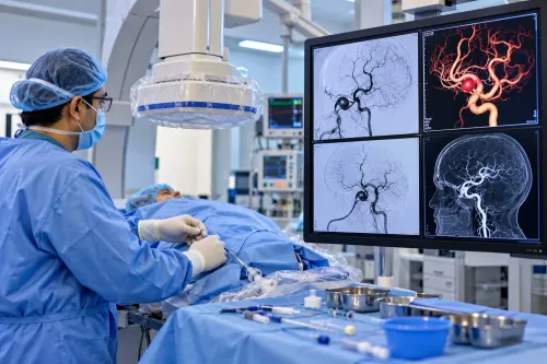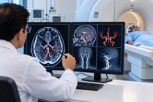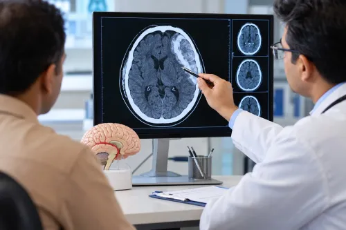
A clot in the brain’s draining veins can obstruct venous outflow, raise intracranial pressure and cause venous infarction or bleeding. Families will learn which symptoms require emergency care, how Pune hospitals confirm the diagnosis, what treatment involves, and how to compare services without assuming that every abnormal scan requires a procedure.
Key takeaways
- Call emergency services for seizure, fainting, weakness, confusion or sudden severe headache.
- Ask whether CT venography or MR venography is needed after a normal plain CT.
- Compare hospitals by neurointervention, neurocritical care and 24-hour imaging access.
- Plan medication reviews, symptom monitoring and follow-up imaging before discharge.
How a brain-vein clot affects the brain and when symptoms are an emergency
A cerebral venous thrombosis (CVST) blocks blood from draining through the brain’s veins or venous sinuses. Back pressure can raise intracranial pressure and cause venous infarction, where brain tissue is injured, or haemorrhage, where damaged vessels bleed. The practical effects include:
- A new or worsening headache; a severe headache requires immediate hospital assessment.
- A seizure, which requires immediate assessment even if it stops.
- Blurred or double vision; rapidly worsening vision is an emergency.
- Weakness, especially sudden weakness on one side; go straight to hospital.
- Confusion or reduced alertness.
- Fainting or loss of consciousness; loss of consciousness is an emergency.
CVST is not the same as an ischemic stroke caused by an artery-blocking clot. The blocked vessel differs, but both conditions can produce weakness, speech or vision problems, seizures and reduced consciousness. A normal early neurological examination does not exclude CVST, particularly when headache or visual symptoms are evolving.
Call emergency services or go to the nearest emergency department for a seizure, sudden weakness, loss of consciousness, severe headache or rapidly worsening vision. Do not wait for a routine neurology appointment; hospital assessment is needed to check for raised intracranial pressure, venous infarction or bleeding.
How Pune hospitals confirm CVST when a plain CT scan is normal
A neurological examination followed by brain CT starts the assessment, but CT alone cannot exclude CVST. A non-contrast CT can look normal or show indirect signs such as swelling, venous infarction or bleeding.
The decisive tests map blood flow in the brain’s draining system:
- CT venography shows the blocked cerebral venous sinus or draining vein after contrast.
- MRI brain identifies venous infarction, swelling and haemorrhage.
- MR venography maps absent or reduced flow in the affected sinus or vein.
| Condition | Clues that overlap with CVST | What helps separate it |
|---|---|---|
| Migraine | Headache, nausea, visual symptoms | New, persistent or progressive headache needs venous imaging |
| Meningitis | Headache, fever, confusion | Fever and cerebrospinal-fluid findings support infection |
| Subarachnoid haemorrhage | Sudden severe headache, vomiting | CT and vascular imaging detect blood or its source |
| Cerebral aneurysm | Headache or neurological deficits | CT angiography identifies an arterial outpouching |
| Subdural haemorrhage | Headache, weakness, reduced alertness | CT shows blood beneath the dura |
Doctors should assess for raised intracranial pressure before considering lumbar puncture. A normal lumbar puncture or initial CT does not rule out CVST when suspicion remains high. This is why cvst treatment in pune begins with the right venous scan rather than reassurance from a normal plain CT.
What prompt medical treatment can prevent clot extension and further brain injury
Once a hospital confirms CVST, admission allows doctors to watch for neurological deterioration, seizures, worsening vision and reduced alertness. Prompt brain vein clot treatment in Pune aims to prevent clot extension, protect threatened brain tissue and support venous recanalisation; it does not instantly dissolve the clot or guarantee recovery.
- Start therapeutic anticoagulation, usually low-molecular-weight heparin. Use unfractionated heparin when severe kidney failure, a likely urgent procedure or rapid reversibility makes it preferable.
- Continue anticoagulation even when venous infarction has caused intracranial haemorrhage, unless the treating team identifies a specific contraindication. It treats the actively thrombosed drainage system and reduces further extension; it does not erase existing bleeding.
- Monitor neurological status, seizures, intracranial pressure and vision. Treat seizures and raised pressure promptly, with measures such as carefully selected fluids, head positioning and specialist therapy.
- Correct dehydration, infection and anaemia, then treat the provoking cause, such as pregnancy-related risk, infection, inflammatory disease or an underlying clotting disorder.
Families comparing cerebral venous thrombosis treatment benefits in Pune should ask which monitoring and treatment are available immediately. Never stop heparin or another prescribed anticoagulant because of headache, bruising or fear of bleeding; contact the treating team first.
A sudden seizure, weakness, worsening alertness or severe headache needs urgent reassessment, not a medication change at home.
When advanced intervention is justified and how to compare Pune hospitals
An abnormal scan alone does not justify a catheter procedure. Catheter-directed thrombolysis or endovascular thrombectomy is rescue treatment for selected patients who deteriorate despite anticoagulation, have severe neurological disease or extensive clot burden, or cannot receive adequate anticoagulation. Evidence does not support it as routine first-line care for every scan showing CVST.
A discussion with a cerebral venous thrombosis specialist in Pune, including Dr Santosh Patil-Endovascular Neuro Surgeon Pune, should clarify how an endovascular option fits the wider hospital plan.
Ask whether the recommendation reflects:
- Neurological deterioration or severe symptoms, not imaging alone
- Haemorrhage, venous infarction, clot location and clot extent
- Raised intracranial pressure and the risk of vision loss
- Contraindications to anticoagulation or failure of adequate treatment
- The centre’s ability to provide rescue intervention and neurocritical care
Compare hospitals by asking for these specific capabilities:
| Capability | Question to ask | Why it matters |
|---|---|---|
| Urgent venous imaging | Can you perform CT venography or MR venography without delay? | Confirms clot location and extent |
| Acute monitoring | Is therapeutic anticoagulation, seizure monitoring and intracranial-pressure management available? | Treats the active disease and its complications |
| Specialist backup | Are neurointerventional, neurosurgical, ophthalmology, haematology and obstetric teams coordinated? | Covers procedures, pressure-related vision problems, clotting disorders and pregnancy |
Interventional neuroradiology alone does not mean thrombectomy is appropriate. Good CVST treatment in Pune combines emergency neurology, anticoagulation, monitoring and escalation when the patient’s course—not the scan alone—makes rescue treatment necessary.
Related service
Cerebral Venous Thrombosis What is Cerebral Venous Thrombosis- Cerebral venous thrombosis (CVT) is a rare but serious condition characterized by the formation of blood clots... View service → |
What recovery planning looks like after the acute clot treatment
After stabilisation, families usually leave with an anticoagulation plan, warning signs to report and scheduled review. One of the cerebral venous thrombosis treatment benefits in Pune is coordinated planning beyond the hospital stay; anticoagulation prevents extension but does not instantly dissolve the clot or guarantee recovery.
Treatment duration is individualised rather than fixed automatically at three months:
- Major transient trigger, such as surgery or infection: about 3–6 months.
- Unprovoked CVST: about 6–12 months.
- Recurrent thrombosis or persistent high-risk thrombophilia: longer treatment, sometimes indefinitely.
A direct oral anticoagulant suits selected adults after the acute phase. Pregnancy, breastfeeding, severe kidney or liver disease, antiphospholipid syndrome, drug interactions and very low or high body weight can change the choice. Warfarin remains important for some patients with antiphospholipid syndrome, so do not choose by cost or convenience alone.
During pregnancy, therapeutic low-molecular-weight heparin is generally preferred. The team must plan doses around delivery and neuraxial anaesthesia, then decide how anticoagulation fits breastfeeding and postpartum risk.
Follow-up covers seizures, persistent headache, papilledema, visual loss, cognition, mood, motor disability and return to work. A continuing headache does not automatically prove clot progression; doctors compare symptoms with examination findings and repeat imaging before changing treatment.
Frequently asked questions
What symptoms make cerebral venous thrombosis an emergency?
Seek emergency care for a sudden severe headache, seizure, fainting, confusion, new weakness, speech difficulty, vision loss or reduced alertness.
Can a plain CT scan miss cerebral venous thrombosis?
Yes. A normal plain CT does not exclude CVST. CT venography or MR venography can show blocked cerebral veins and venous sinuses.
What does prompt CVST treatment aim to prevent?
Treatment aims to stop clot extension, restore venous drainage and limit pressure, venous infarction, haemorrhage and further brain injury.
When is advanced intervention considered for CVST?
A neurovascular team may consider endovascular treatment when the patient deteriorates or remains critically ill despite appropriate medical treatment.
What should recovery planning include after CVST treatment?
Before discharge, plan anticoagulation review, neurological follow-up, rehabilitation if needed, warning-sign instructions and follow-up imaging.
Keywords