A subdural bleed in an older adult may begin with a minor fall and look like forgetfulness, imbalance, or unusual sleepiness rather than a dramatic emergency. You will learn which symptoms need immediate hospital assessment, how doctors choose between monitoring, drainage, and embolisation, and what benefits and trade-offs to discuss with the treating team.
Key takeaways
- Treat new confusion, weakness, seizures, or severe drowsiness as urgent warning signs.
- Ask how CT findings, bleed size, symptoms, and medicines affect treatment timing.
- Compare observation, burr-hole drainage, craniotomy, and embolisation by risks and recovery needs.
- Choose a Pune hospital with neurovascular, neurosurgical, imaging, and rehabilitation support.
How a subdural bleed can look like ageing or dementia
A subdural bleed can look like dementia because its symptoms may build slowly: headache, confusion, drowsiness, memory loss, personality change, imbalance, repeated falls, weakness, speech difficulty, or seizures. Brain shrinkage in older adults stretches the bridging veins between the brain and its outer covering, so a seemingly minor fall can tear them.
Warfarin, apixaban, rivaroxaban, aspirin, and clopidogrel can increase bleeding or prolong it. Recognising this pattern is central to understanding how subdural hemorrhage treatment benefits older adults in Pune.
| Type | Timing | Common presentation |
|---|---|---|
| Acute | Minutes to hours after a significant injury | Severe headache, vomiting, drowsiness, weakness, speech difficulty, seizure, or loss of consciousness |
| Subacute | About 2 to 14 days after injury | Worsening headache, confusion, imbalance, falls, drowsiness, or one-sided weakness |
| Chronic | More than 2 weeks later, sometimes after a minor or forgotten fall | Memory or personality change, reduced walking ability, lethargy, speech problems, headache, or repeated falls |
Do not arrange a routine appointment if any emergency sign appears. Seek immediate hospital assessment in Pune for:
- Worsening alertness or loss of consciousness
- Repeated vomiting
- New one-sided weakness or speech difficulty
- A seizure
- Unequal pupils
How doctors confirm the bleed and decide whether treatment is urgent
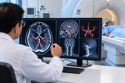
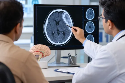
A non-contrast CT head scan usually makes the first treatment decision because it quickly shows blood, collection thickness, midline shift, and mass effect. MRI adds detail when CT is unclear or the collection is slow-developing, organised, or difficult to distinguish from another cause of decline.
Clinicians then check:
- Alertness, confusion, speech, strength, pupils, walking ability, and seizure activity.
- Whether symptoms are worsening, persistent, or newly appearing after a period of stability.
- Frailty, baseline cognition, mobility, fall risk, kidney function, heart and lung disease, and other illnesses.
- Whether warfarin, apixaban, rivaroxaban, aspirin, clopidogrel, or another antithrombotic needs reversal or interruption.
A small, stable, minimally symptomatic collection can suit observation with neurological checks and scheduled repeat CT scans. Deterioration, substantial midline shift, dangerous pressure, or persistent disabling symptoms calls for urgent neurosurgical treatment.
Age alone does not decide the balance: a fit 85-year-old may benefit from treatment, while advanced dementia or severe frailty may limit recovery even after technically successful evacuation.
For elderly subdural hemorrhage treatment in Pune, ask whether the plan covers reversal, interruption, and restart of antithrombotic medicine. Restart timing must match the bleeding pattern and the competing risk of stroke, heart attack, or stent thrombosis; no single day is safe for everyone.
What observation, drainage, and craniotomy each offer
Observation avoids an operation, but it is safe only with scheduled neurological checks, repeat CT imaging, and clear instructions for deterioration. It fits a small, stable, minimally symptomatic collection; worsening alertness, new weakness, increasing headache, or dangerous mass effect changes the decision.
| Option | What it offers | Main trade-off |
|---|---|---|
| Observation | No surgical wound or anaesthesia; repeat scans track change | Pressure can worsen between reviews, so deterioration needs urgent drainage |
| Burr-hole or twist-drill drainage | Burr-hole evacuation with a temporary drain usually decompresses a symptomatic chronic collection faster than observation; twist-drill drainage uses less surgical exposure in selected cases | Infection, acute rebleeding, seizures, tension pneumocephalus, venous thromboembolism, and recurrence remain possible |
| Craniotomy | Wider access for an organised, recurrent, or complex clot | More operative time, tissue exposure, anaesthetic burden, and recovery demands |
Medication review belongs in every pathway. Reversal or temporary interruption of warfarin, apixaban, rivaroxaban, aspirin, or a P2Y12 inhibitor can reduce further bleeding, but stopping treatment can raise the risk of stroke, heart attack, or stent thrombosis. The restart date must match the bleed, the procedure, and the reason for antithrombotic treatment.
For subdural bleeding care in Pune, ask how the team will manage these complications:
- Infection or acute rebleeding after drainage
- Seizures or tension pneumocephalus
- Venous thromboembolism during reduced mobility
- Recurrence requiring repeat imaging or treatment
A drain does not end monitoring. Follow-up CT and neurological review detect residual or recurrent blood, while mobility support and fall prevention reduce avoidable setbacks.
Where middle meningeal artery embolisation may benefit selected patients
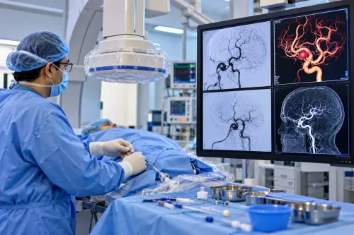
Middle meningeal artery embolisation (MMAE) blocks arterial branches that supply the fragile membrane around some chronic subdural collections. By reducing this membrane’s blood flow, it may lower recurrence after drainage or help selected patients avoid, delay, or limit surgical exposure. It does not rapidly remove a large clot.
| Option | Potential benefit | Boundary |
|---|---|---|
| Drainage alone | Quickly reduces pressure by removing fluid or clot | Does not directly treat the membrane’s blood supply |
| Drainage plus MMAE | EMBOLISE found 4.1% recurrence or progression requiring repeat surgery, versus 11.3% with surgery alone | Evidence supports selected use, not treatment for every patient |
| MMAE alone | Avoids an operation in carefully selected chronic or recurrent cases | Cannot replace emergency decompression or remove severe mass effect |
MAGIC-MT reported a neutral primary result, so the evidence is promising rather than universal. Doctors still match the approach to CT findings, neurological status, recurrence pattern, frailty, and the angiogram.
Risks include contrast reaction, kidney injury, arterial or groin-access bleeding, ischemic stroke, retinal injury, cranial-nerve injury, and non-target embolisation through connections to the ophthalmic or intracranial circulation. Angiography identifies these pathways but cannot make risk zero.
For subdural bleeding care in pune, Dr Santosh Patil-Endovascular Neuro Surgeon Pune should coordinate with neurosurgery, anaesthesia, CT, and emergency decompression services. If you consult a subdural hematoma specialist in pune, ask whether the plan is standalone MMAE, drainage, or both, and what happens if consciousness worsens or the collection returns.
Related service
Subdural Hemorrhage What is Subdural Hemorrhage ? Subdural hemorrhage refers to the accumulation of blood between the dura mater and the arachnoid mater layers of the... View service → |
How to compare hospitals, recovery plans, and specialist advice in Pune
A hospital for elderly subdural hemorrhage treatment in Pune should offer urgent CT, neurosurgery, neurocritical care, anaesthesia, and experienced interventional neuroradiology when embolisation is considered. Confirm after-hours surgical backup; a clinic label cannot replace immediate decompression if the patient deteriorates.
| Option | What it means | Questions to ask |
|---|---|---|
| Observation | Repeat neurological checks and CT scans | Why is observation safe, and when is the next scan? |
| Drainage | Burr-hole or twist-drill evacuation, often with a temporary drain | Is the collection chronic, causing mass effect, and how will the drain be managed? |
| Embolisation | Middle meningeal artery treatment targeting chronic membrane blood supply | Is it standalone or adjunctive, what angiographic risks exist, and what happens if symptoms worsen? |
| Both | Drainage for pressure plus embolisation to reduce selected recurrence risk | Why does this patient need both rather than one procedure? |
- Is the collection acute, subacute, or chronic, and is there midline shift or dangerous mass effect?
- What finding supports observation or intervention?
- When will warfarin, apixaban, rivaroxaban, aspirin, or clopidogrel restart?
- What rehabilitation, mobility, nutrition, and fall-prevention plan begins after discharge?
- How will recurrence be detected and treated?
A consultation with a subdural hematoma specialist in Pune should identify the clinician as a neurosurgeon, neurologist, or interventional neuroradiologist and explain their coordination.
Frailty, baseline cognition, mobility, renal function, goals of care, and the actual cause of decline affect benefit as much as the CT; treatment may not restore independence in advanced dementia or severe frailty.
Frequently asked questions
How can a subdural bleed look like ageing or dementia?
A slow bleed can cause confusion, memory loss, personality change, drowsiness, imbalance, falls, weakness, speech difficulty, or seizures. New or worsening symptoms need medical assessment rather than being attributed to ageing.
How do doctors confirm a subdural bleed and decide whether treatment is urgent?
Doctors use a neurological examination and brain imaging, usually a CT scan. Urgency depends on symptoms, the collection’s size and pressure effect, its location, progression, and medicines such as anticoagulants.
What do observation, drainage, and craniotomy each offer?
Observation uses repeat examinations and scans when the bleed is small, stable, and causing limited symptoms. Burr-hole drainage removes suitable collections through small openings. Craniotomy provides wider access when the clot is organised, thick, recurrent, or difficult to drain.
Where might middle meningeal artery embolisation benefit selected patients?
Middle meningeal artery embolisation reduces blood flow to vessels feeding a chronic subdural collection. A specialist may consider it for selected patients with recurrence risk or alongside drainage; it does not replace urgent decompression when brain pressure is dangerous.
How should you compare hospitals and specialist advice in Pune?
Ask about neurosurgical and endovascular expertise, 24-hour CT access, intensive care, anticoagulant management, embolisation availability, follow-up scans, rehabilitation, and who coordinates recovery at home.
Keywords