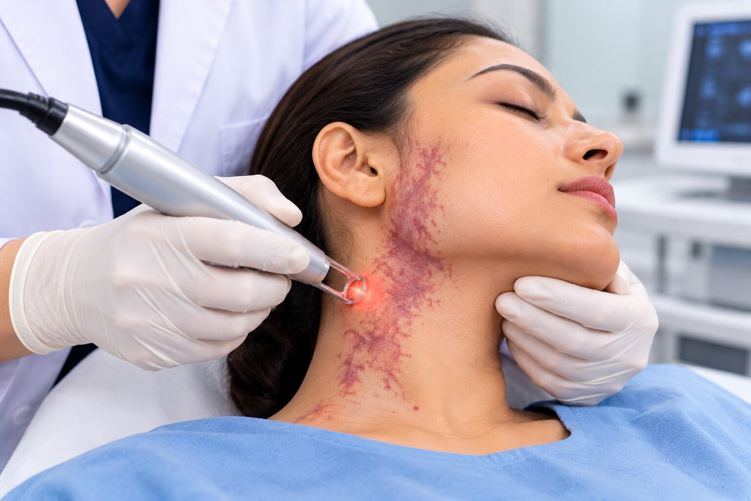
A visible or enlarging mark, lump, or swelling in the face, neck, mouth, or scalp does not automatically require an operation, but pain, bleeding, functional problems, and high-flow features need careful assessment. By the end, you will know which symptoms and lesion types justify specialist review, which tests clarify the diagnosis, and when embolization, sclerotherapy, surgery, or monitoring may be appropriate.
Key takeaways
- Seek assessment for growth, bleeding, pain, ulceration, pulsation, or breathing difficulty.
- MRI and Doppler ultrasound help define the lesion before treatment decisions.
- A multidisciplinary team coordinates imaging, embolization, surgery, and follow-up.
- Embolization can reduce blood flow or bleeding before selected procedures.
Which symptoms mean a head and neck vascular malformation needs assessment?
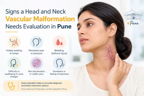
Assessment is warranted when a birthmark or swelling enlarges, feels compressible or pulsates, or causes recurrent bleeding, pain, ulceration, infection, facial asymmetry, breathing or swallowing difficulty, speech changes, hearing or vision symptoms, or progressive limitation in daily activities. These are the people who may need head and neck vascular malformation treatment in Pune.
| Situation | Warning signs | What to do |
|---|---|---|
| Stable lesion | Incidental finding with no pain, bleeding, growth, or functional effect | Arrange routine specialist assessment; monitoring may be safer than immediate treatment |
| Prompt review | Threat to the airway, nerves, eyes, major vessels, or daily function; progressive enlargement or repeated infection | Seek specialist evaluation soon |
| Emergency | Uncontrolled bleeding, sudden painful enlargement, breathing difficulty, airway compression, new weakness or speech difficulty, loss of vision, or rapidly developing neurological symptoms | Go to emergency care immediately |
A stable, symptom-free malformation does not automatically need an operation. Observation, compression, medicines, sclerotherapy, embolization, surgery, or combined treatment depend on its flow pattern, anatomy, growth, and the risk of leaving it untreated.
For children, arrange assessment when feeding, sleep, development, speech, hearing, vision, breathing, or daily function is affected, or when bleeding or infection recurs. Treatment timing balances those risks against anaesthesia, radiation, post-procedure swelling, and the possibility of several procedures.
How can you tell a vascular malformation from another lump or birthmark?
A lesion’s flow and behaviour offer more useful clues than colour alone, but only examination and imaging can confirm the diagnosis.
| Feature | Practical clue | What it suggests |
|---|---|---|
| Venous malformation | Soft, blue or purple, compressible; enlarges when the head is lowered or during straining | Low-flow venous component |
| Lymphatic malformation | Soft or cystic; swelling increases after infection, inflammation, or bleeding | Low-flow lymphatic component |
| Capillary malformation | Flat red, pink, or purple patch without a deep lump | Superficial capillary change |
| Arteriovenous malformation or fistula | Warmth, visible pulsation, rapid filling, a palpable thrill, or an audible bruit | High-flow arterial connection |
| Mixed malformation | Features of two or more types, such as a compressible mass with warmth or pulsation | Combined vascular channels |
| Infantile haemangioma | Usually appears after birth, grows during an early proliferative phase, then gradually involutes | Different condition from a structural malformation |
| Tumour, lymph node, or cyst | A solid mass, discrete node, or defined fluid-filled lump that does not follow vascular filling patterns | Non-malformation cause requiring assessment |
| Ordinary varicose vein | A superficial, rope-like vein following a typical venous pathway | Local venous disease, not necessarily a malformation |
Seek review for a lesion present since childhood that becomes clearer with growth, or one that enlarges after puberty, pregnancy, hormonal change, infection, or injury. Sudden change, pain, bleeding, ulceration, or diagnostic uncertainty also warrants assessment. Appearance alone cannot establish flow type or safely exclude a tumour.
Which tests show whether treatment is safe and useful?
A safe plan starts by identifying flow, depth, extent, and nearby structures—not by judging appearance alone. Physical examination checks colour, warmth, compressibility, pulsation, bruit, skin changes, neurological function, airway involvement, and changes with position or the Valsalva manoeuvre.
| Investigation | Question it answers | When it applies |
|---|---|---|
| Physical examination | Does the lesion behave like a low-flow or high-flow abnormality, and is it affecting nerves, skin, or the airway? | Every assessment |
| Duplex Doppler ultrasound | Is flow slow and venous, or fast and arterial? | Superficial lesions; no radiation |
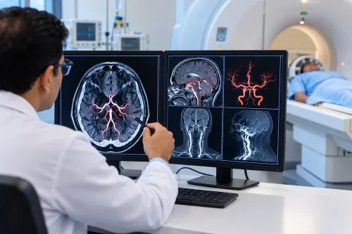
| Contrast MRI with dynamic vascular sequences | What is the lesion’s volume and relationship to muscles, nerves, airway, bone, orbit, and brain? | Deep, extensive, or anatomically complex lesions |
| CT angiography or MR angiography | What are the arterial anatomy, bone changes, and vascular connections? | When detailed cross-sectional mapping is needed |
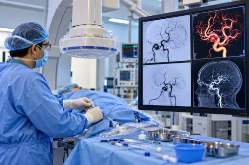
| Catheter angiography | Where are the feeding arteries, AVM nidus, venous drainage, and safest treatment access? | Suspected arteriovenous malformation or fistula when embolization is considered |
Before vascular malformation treatment in Pune, prepare the team with:
- Prior scans and discs, biopsy reports, procedure and medication records, and photographs showing change over time.
- A list of anticoagulant or antiplatelet medicines.
- Information about pregnancy or possible pregnancy, kidney disease, contrast reactions, allergies, and bleeding or clotting disorders.
These details can change the imaging method, contrast plan, anaesthesia, medication management, or procedure timing.
Who should coordinate treatment, and when does embolization help?
Choose a head neck vascular malformation specialist in Pune who evaluates both low-flow and high-flow lesions and coordinates imaging, intervention, surgery, anaesthesia, and follow-up. A clinician experienced with ordinary varicose veins or routine aneurysms may not have equivalent expertise in facial, skull-base, airway, or intracranial malformations.
| Specialist | Main contribution |
|---|---|
| Interventional neuroradiologist or interventional radiologist | High-flow AVM, fistula, skull-base lesion, angiography, or catheter treatment |
| ENT or head-and-neck surgeon | Airway, throat, larynx, swallowing, and surgical access |
| Plastic surgeon | Reconstruction, facial symmetry, and function |
| Vascular surgeon | Selected extracranial arterial or venous anatomy |
| Dermatologist | Superficial capillary lesions |
| Paediatrician | Coordination of children’s care |
| Neurosurgeon | Brain, dura, or critical intracranial vessels |
Embolization helps most when imaging identifies a safely reachable treatment target. In an AVM, the target is the nidus—the abnormal vascular tangle—not merely a proximal feeding artery; blocking only the feeder can recruit new vessels and allow recurrence. Treatment may be staged, combined with surgery, or used alone when complete nidus access is safe.
For a venous or lymphatic malformation, “embolization” often means image-guided percutaneous sclerotherapy: a needle delivers a sclerosant into the abnormal spaces. Lesion volume, drainage, and proximity to the eyes, skin, nerves, airway, and brain determine the number and type of procedures.
Dr Santosh Patil-Endovascular Neuro Surgeon Pune can contribute when skull-base, intracranial, or catheter-based neurovascular assessment is relevant to the team’s plan.
Related service
Head And Neck Vascular Malformations Head And Neck Vascular Malformations What is Head & Neck Vascular Malformations- Head & Neck Vascular Malformations encompass a diverse group of anomalies involving abnormal blood... View service → |
What benefits, risks, and follow-up should you expect?
Treatment earns its burden when it reduces bleeding, pain, infection, swelling, disfigurement, airway or swallowing problems, neurological risk, or progressive tissue damage. A visible lesion alone is not a reason for intervention; a stable, symptom-free malformation may be monitored.
Options include observation, compression, supportive care, selected medicines, image-guided sclerotherapy, embolization, surgery, or a combination. High-flow AVMs need treatment directed at the nidus, while symptomatic low-flow venous or lymphatic lesions are more often considered for sclerotherapy or surgery.
Head-and-neck embolization can cause non-target embolization, skin or mucosal necrosis, cranial-nerve injury, stroke, bleeding, infection, contrast reaction, kidney injury in susceptible patients, and radiation exposure. Post-treatment swelling can temporarily worsen breathing or swallowing. Lesions involving the tongue, pharynx, larynx, or airway require an explicit airway plan.
Before agreeing to treatment, ask:
- Does the procedure target the AVM nidus rather than only a feeder?
- How will normal arteries and venous drainage be protected?
- Is staged treatment likely, and what happens if the lesion remains?
- How will recurrence or residual disease be checked?
A technically successful session does not always end care. Examination, MRI, or repeat angiography may detect residual or recurrent AVM; low-flow lesions can refill or become symptomatic. Children need longer review because growth changes the lesion and surrounding anatomy.
Frequently asked questions
Which symptoms mean a head and neck vascular malformation needs assessment?
Arrange assessment for enlargement, compressibility, pulsation, recurrent bleeding, pain, ulceration, infection, facial asymmetry, breathing or swallowing difficulty, speech changes, hearing or vision symptoms, or worsening limits on daily activities.
How can you tell a vascular malformation from another lump or birthmark?
A clinician assesses its colour, temperature, compressibility, pulsation, change with position or straining, growth pattern, and relationship to nearby structures. Doppler ultrasound and MRI help distinguish blood-flow lesions from cysts, tumours, lymph nodes, and ordinary pigmented birthmarks.
Which tests show whether treatment is safe and useful?
Doppler ultrasound evaluates flow and vessel connections. MRI with contrast maps the lesion and its depth. CT or CT angiography helps assess bone and airway relationships, while catheter angiography provides a detailed treatment map when embolization is being considered.
Who should coordinate treatment, and when does embolization help?
A vascular anomaly team may include an endovascular neurointerventional specialist, head and neck surgeon, radiologist, anaesthetist, and other specialists based on the affected structures. Embolization helps selected high-flow lesions, active bleeding, or lesions requiring blood-flow reduction before surgery.
What benefits, risks, and follow-up should you expect?
Treatment aims to control bleeding, pain, growth, disfigurement, functional problems, or airway and swallowing symptoms. Risks include bleeding, infection, skin or tissue injury, nerve damage, unintended vessel blockage, contrast reactions, and recurrence. Follow-up uses clinical review and repeat imaging when needed.
Keywords